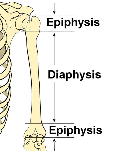
Long Bone Diagram : Cybersurgeons / Long, short, flat, irregular and sesamoid.long bones, especially the femur and tibia, are subjected to most of the load during daily activities and they are crucial for skeletal mobility.they grow primarily by elongation of the diaphysis, with an epiphysis at each end of the growing bone.
Long Bone Diagram : Cybersurgeons / Long, short, flat, irregular and sesamoid.long bones, especially the femur and tibia, are subjected to most of the load during daily activities and they are crucial for skeletal mobility.they grow primarily by elongation of the diaphysis, with an epiphysis at each end of the growing bone.. Related posts of diagram of a long bone anatomy anatomy kidney function diagram. A long bone has two main regions: Plates of cartilage, also known as growth plates which allow the long bones to grow during childhood. In the diagram of the femur (thigh bone) on the right, notice that the shapes of the upper and lower epiphyses are different. Learn vocabulary, terms, and more with flashcards, games, and other study tools.
The femur, or thighbone, is the longest and largest bone in the human body. Area between the diaphysis and epiphysis at both ends of the bone. The ends of long bones are called epiphyses. This long bone connects with the knee at one end and the ankle at the other. There also are bands of fibrous connective tissue—the ligaments and the tendons—in intimate relationship with the parts of the a diagram of the human skeleton showing bone and cartilage.

Click on the tags below to find other quizzes on the same subject.
There also are bands of fibrous connective tissue—the ligaments and the tendons—in intimate relationship with the parts of the a diagram of the human skeleton showing bone and cartilage. Long bone diagram|long bone diagram|how to draw long bonehi friends, in this video we will learn how to draw diagram of long bone#longbonediagram. Click on the tags below to find other quizzes on the same subject. The diaphysis and the epiphysis (figure 6.3.1). Used figure 6.2 in book. Structure of the long bone with pictures learn with flashcards, games, and more — for free. The femur, or thighbone, is the longest and largest bone in the human body. The outside of the flat bone consists of a layer of connective tissue called the periosteum. The diaphysis is the tubular shaft that runs between the proximal and distal ends of the bone. Learn long bone diagram with free interactive flashcards. This long bone connects with the knee at one end and the ankle at the other. Related posts of cross section of a long bone bone structure birds. A typical long bone shows the gross anatomical characteristics of bone.
The structure of a long bone consists of several sections:. Related posts of diagram of a long bone anatomy anatomy kidney function diagram. There is a printable worksheet available for download here so you can take the quiz with pen and paper. The outside of the flat bone consists of a layer of connective tissue called the periosteum. Inside the diaphysis is the medullary cavity, which is filled with yellow bone marrow in an adult.

The ends of long bones are called epiphyses.
A long bone has two main regions: Parts of long bone (applies to other bones too). A typical long bone shows the gross anatomical characteristics of bone. Start learning with our skeleton diagrams, bone labeling exercises and skeletal system quizzes! Smartdraw includes 1000s of professional healthcare and anatomy chart templates that you can modify and make your own. The femur, or thighbone, is the longest and largest bone in the human body. Bones in the hand and wrist right hand 12 photos of the bones in the hand and wrist right hand , bone Diagram of a long bone, diagram of long bone structure, diagram of long bone to label, structure of a long bone diagram quiz, unlabeled diagram of a long bone, bone. Related posts of cross section of a long bone bone structure birds. Plates of cartilage, also known as growth plates which allow the long bones to grow during childhood. Each end is specially designed to fit the bone or bones it attaches to. The diaphysis is the tubular shaft that runs between the proximal and distal ends of the bone. Area between the diaphysis and epiphysis at both ends of the bone.
Parts of long bone (applies to other bones too). The diaphysis is the hollow, tubular shaft that runs between the proximal and distal ends of the bone. A long bone is a bone that has greater length than width. This is an online quiz called long bone diagram there is a printable worksheet available for download here so you can take the quiz with pen and paper. The largest bone in the human body is the thighbone or femur, and the smallest is the stapes in the middle ear, which are just 3 millimeters (mm) long.

Use the text and the diagram to help you to answer the questions.
This long bone connects with the knee at one end and the ankle at the other. Use our interactive diagram to explore the different parts of the skeletal system. Bones of the hand diagram 12 photos of the bones of the hand diagram bones in the hand diagram. The ends of long bones are called epiphyses. Long bones have a thick outside layer of compact bone and an inner medullary cavity containing bone marrow. Related posts of cross section of a long bone bone structure birds. It is also known as the calf bone, as it. Related posts of diagram of a long bone anatomy anatomy kidney function diagram. The diaphysis is the hollow, tubular shaft that runs between the proximal and distal ends of the bone. Once we stop growing (between 18. The ulna is the second long bone of the forearm, found on the pinky finger side. Long bones are those that are longer than they are wide. Related posts of 3d diagram of long bone bones of the hand diagram.


Komentar
Posting Komentar